What is Capsule Endoscopy? How And Why Is It Done?

Click to rate this post!
[Total: 1 Average: 5]
Capsule endoscopy is a stylish video capsule designed in the size of a large pill, with its own light source and camera.

Capsule Endoscopy

kapsül endoskopi nasıl kullanılırAs is known, the digestive system is in the form of a long canal extending from the mouth to the anus. While it was possible to examine the esophagus, stomach, duodenum and large intestine, which constitute the beginning and end parts of this canal, by endoscopic methods such as gastroscopy and colonoscopy, it was not possible to examine the middle parts consisting of three parts called the duodenum, jejunum and ileum.

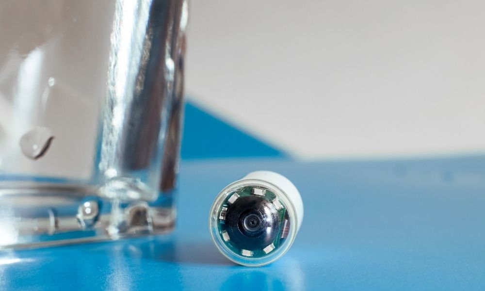
Capsule endoscopy, a method developed for this purpose, is a large pill-sized video capsule with its own light source and camera. You may also hear about capsule endoscopy from your doctor or other healthcare professionals by different names such as small bowel endoscopy, capsule enteroscopy or wireless endoscopy. The system consists of a 12mmx26mm diameter capsule and a recorder, the size of a small handheld radio, which is attached to your waist with a belt, which receives images and sends these images with radiofrequency waves. After recording, the data in the recorder is transferred to a computer with a special program and the recorded images are examined. Wireless capsule endoscopy (wireless capsule endoscopy) is also called ‘pill glass’ or M2A (Mouth to anus).
kapsul endoskopi nasil yapilir

During the procedure, the patient can continue his daily activities and take food. The recording time is about 6-8 hours. Pathological lesion that may cause bleeding can be found in 65-75% of patients with unexplained digestive system bleeding, and 25-35% of patients may not get results with this method.

It is painless and does not require sedation, food can be eaten during the procedure. The main disadvantages are the inability to intervene in the lesion, the inability to take a tissue sample (biopsy), the inability to examine the lesion by imaging for a long time, and the inability to always obtain a quality image. In 0.7-2% of patients, the capsule may remain in the intestines and not be excreted with stool, and surgical intervention may be required to remove it.

It may be inconvenient to use in patients with swallowing difficulties, pregnant women, and patients with cardiac pacemaker. It cannot be used in the presence of stenosis and large diverticulum in the intestines.

Why is Capsule Endoscopy Performed?

kapsül endoskopi nedirWhile it is possible to easily reach and examine the upper (oesophagus, stomach and duodenum) and lower (last 15-20 cm part of the large intestine and small intestine) parts of the digestive system with gastroscopy and colonoscopy, it is possible to examine these parts of the small intestine with a length of approximately 4.5-5.5 m.

It is difficult to visualize the inner surface with these methods. For this purpose, a device called capsule endoscopy has been developed. Although capsule endoscopy is a method developed for the examination of the small intestines, in some cases it is possible to see lesions in the esophagus, stomach and large intestines. With this method, it is possible to reveal the cause of small intestinal bleeding, and to have information about other diseases of the small intestine (polyp, Crohn’s disease, ulcers, absorption disorders and small intestine tumors, etc.).

How is Capsule Endoscopy Performed?

Inform your doctor if you have a drug that you use regularly. Some drugs, such as iron, bismuth, and antidiarrheal drugs, may prevent the device from getting images or progressing in the intestine. The daily doses of the drugs you use may need to be adjusted again before the test.

If you have a disease such as difficulty in swallowing, chronic inflammatory bowel disease such as Crohn’s disease, chronic stomach or duodenal (duodenal) ulcer, if you are aware of the presence of large diverticula in your esophagus or intestines due to a previous examination, If you have previously had intestinal obstruction, intestinal adhesions or abdominal surgery, if you have a pacemaker, you should definitely inform your doctor about these issues.

If necessary, your doctor may want you to have a small bowel X-ray before the capsule endoscopy application in order to determine whether there is a stenosis in your intestines that may prevent the passage of the capsule.

kapsül endoskopi

In order for the test to be done well and reliably, nothing should be eaten or drunk until at least 12 hours before the test. Your doctor will inform you when you can start eating. Shopping centers etc. Since the magnetic field at the electronic security doors at the entrances of the places may interact with the capsule, care should be taken not to pass through such doors during the registration.

Before the capsule is swallowed, a recording device (recorder) is attached to your waist with the help of a tape or belt, and then the capsule is swallowed with some water. While the capsule moves in the intestine with the peristaltic movements of the intestines, the video images obtained simultaneously are recorded on the sensing device placed on your belt for 6-8 hours.

Unless your doctor recommends anything else, you can eat water and similar liquids 4 hours after the capsule is swallowed, and a light meal 6 hours later. You should avoid heavy exercise movements such as running and jumping while the procedure is in progress.

At the end of the process, the data in the recorder is transferred to a computer with a special program and evaluated by a doctor trained in this subject. The capsule in the intestines is excreted with feces within 12-48 hours. When it is not seen that the capsule is excreted in the stool, a direct abdominal X-ray can be taken 1-2 weeks after the procedure and it can be checked whether the capsule is expelled or not.

Bir Cevap Yazın

Your email address will not be published.