What is Autoimmune Pancreatitis? Diagnosis and Treatment

Click to rate this post!
[Total: 2 Average: 5]
Autoimmune pancreatitis is a rare form of chronic pancreatitis due to autoimmune etiology. It may occur as a primary pancreatic disease or may progress with other autoimmune diseases (eg primary sclerosing cholangitis, primary biliary cirrhosis, retroperitoneal fibrosis, rheumatoid arthritis, sarcoidosis, Sjögren’s syndrome).

Otoimmün Pankreatit nedirIt is also called autoimmune pancreatitis, sclerosing pancreatitis, nonalcoholic destructive pancreatitis. HISORt diagnostic criteria developed by Mayo Clinic are used in diagnosis.

Although glucocorticoids are used in the treatment (recommended dose: starting prednisone 40 mg/day and stopping the treatment after 4-6 weeks by 5 mg per week), the optimal dose and duration are still unknown.

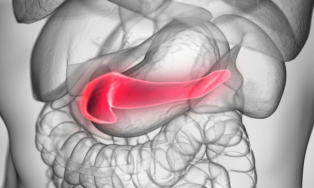
Autoimmune pancreatitis is a form of chronic pancreatitis with distinctive clinical, histological and morphological features that develops due to autoimmune etiology.

This form of chronic pancreatitis was first described by Sarles et al. in 1961, and the term autoimmune pancreatitis was first used by Yoshida et al. in 1995. In 2003, Kamisawa et al. reported that autoimmune pancreatitis is a systemic disease, and the presence of IgG4-positively stained plasma cells in the pancreas and other affected organs (4).

Although the first reported cases in the literature were from Japan, the incidence is increasing all over the world with the increase in detectability. Autoimmune pancreatitis; It may occur as a primary pancreatic disease or may progress with other autoimmune diseases (eg primary sclerosing cholangitis, primary biliary cirrhosis, retroperitoneal fibrosis, rheumatoid arthritis, sarcoidosis, Sjögren’s syndrome).

Otoimmun Pankreatit tedavisi

Autoimmune pancreatitis can also be seen as a focal lesion mimicking carcinoma of the pancreas. Although this condition is most commonly seen in the head of the pancreas, it may also be located in the trunk and tail, or it may be found in the form of multiple lesions.

Terminology?

It is also called autoimmune pancreatitis, sclerosing pancreatitis, nonalcoholic destructive pancreatitis (8,9). Detection of IgG4 positive staining plasma cells in the pancreas or other affected tissues is an important finding supporting autoimmune pancreatitis.

It has been emphasized in the literature that IgG4-related cholangitis is not a separate clinical picture, but a biliary manifestation of autoimmune pancreatitis.

Clinical Findings

  • Moderate abdominal pain, with or without pancreatitis attack,
  • Obstructive jaundice,
  • Diffuse swelling and enlargement especially evident in the head of the pancreas on imaging, mimicking pancreatic carcinoma in the late stage
  • Diffuse irregularity in the pancreatic duct,
  • Increased serum gammaglobulin level, especially IgG4 level,
  • Other autoantibody [such as antinuclear antibody (ANA), rheumatoid factor (RF)] positivity,
  • Cyst or calcification in the pancreas is not often accompanied,
  • Association with other autoimmune diseases (such as Sjögren, sclerosing cholangitis, ulcerative colitis, rheumatoid arthritis),
  • Stenosis in the main bile duct and intrahepatic bile ducts,
  • Presence of advanced fibrosis and lymphoplasmocytic infiltration in pancreatic biopsies,
  • Reduction in pancreatic size and improvement in histological changes with corticosteroid therapy.
Otoimmün Pankreatit belirtileri

Extrapancreatic Findings

  • Inflammatory bowel disease (often ulcerative colitis),
  • Autoimmune sclerosing cholangitis,
  • Nodules, adenopathy and infiltrates in the lung,
  • Sjogren’s syndrome,
  • Retroperitoneal fibrosis,
  • Long segment structures often occur in the bile ducts,
  • Tubulointerstitial nephritis,
  • Autoimmune thyroiditis.


Clinical Profile

Otoimmün Pankreatit tanısıThere are two different histological types of autoimmune pancreatitis and the clinical findings of these two different histological types are also different. Type 1 autoimmune pancreatitis Also called lymphoplasmocytic sclerosing pancreatitis . Diagnosed by microscopic examination at high magnification, by the presence of more than 10 IgG4-positive staining cells and at least two of the following:

  • Periductal lymphoplasmocytic infiltration,
  • Obliterative phlebitis,
  • Acinar fibrosis.
    Type 2 Autoimmune Pancreatitis
    It is called idiopathic duct centric pancreatitis. Neutrophilic infiltration in the pancreatic duct and detection of IgG4 positive staining cells in the pancreatic parenchyma are supportive findings.
  • Patients with type 1 autoimmune pancreatitis are often older and have higher IgG4 levels than type 2 autoimmune pancreatitis, with more relapses in this group.
  • Extrapancreatic autoimmune diseases are more common in type 1 autoimmune pancreatitis.

Diagnosis of Autoimmune Pancreatitis

  • The most important point in the differential diagnosis is autoimmune pancreatitisand pancreatic cancer
  • Pancreatic biopsy is a necessary method for diagnosis, and detection of IgG4 positive plasma cell infiltration in the pancreas and other affected organs are supportive findings.
  • The diagnostic criteria of the Japanese Pancreas Society can be used for diagnosis, and recently the Mayo Clinic HISORt diagnostic criteria are used for diagnosis.

Treatment of Autoimmune Pancreatitis

stroid tedavisi

In autoimmune pancreatitis, glucocorticoids regress clinical findings and prevent complications (17). Steroid therapy usually provides a lasting clinical and radiological remission. It was found that serological findings improved and stenosis in the pancreatic ducts disappeared with steroid treatment. In some series, it has been shown that recurrent pancreatitis, structures or extrapancreatic disease may develop in 10% of patients with steroid therapy.

Prednisone is usually started at a dose of 0.4-0.6 mg/kg/day. After 4-6 weeks of treatment, the dose is tapered to 5 mg per week. Most of the patients are in long-term remission with 2-3 months of corticosteroid treatment, but some patients may require maintenance treatment or relapse with no response to treatment. Time to treatment response can range from two weeks to four months. During treatment, patients should be evaluated with IgG4 level and other biochemical and radiological imaging methods.

Although there is limited data, immunomodulatory drugs can be used in cases with treatment failure or relapse under steroid therapy.

In a study conducted on 13 patients with autoimmune pancreatitis, 10 patients were treated with azathioprine 2 mg/kg/day, remission was achieved in seven patients with azathioprine monotherapy, and the follow-up period was 14 months (19).

Treatment success was achieved with rituximab (monoclonal antibody) treatment in two patients with autoimmune pancreatitis who did not respond to glucocorticoid and 6-mercaptopurine treatment (20). However, further studies on these treatments are needed. The most important uncertainty in treatment is how long the treatment period will continue.

Bir Cevap Yazın

Your email address will not be published.